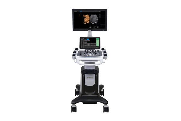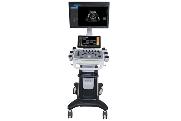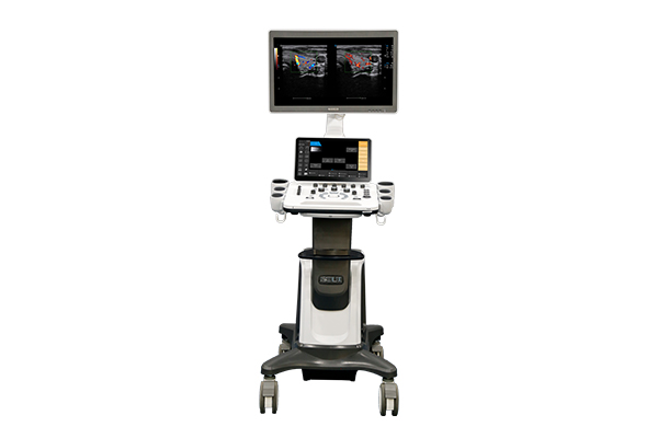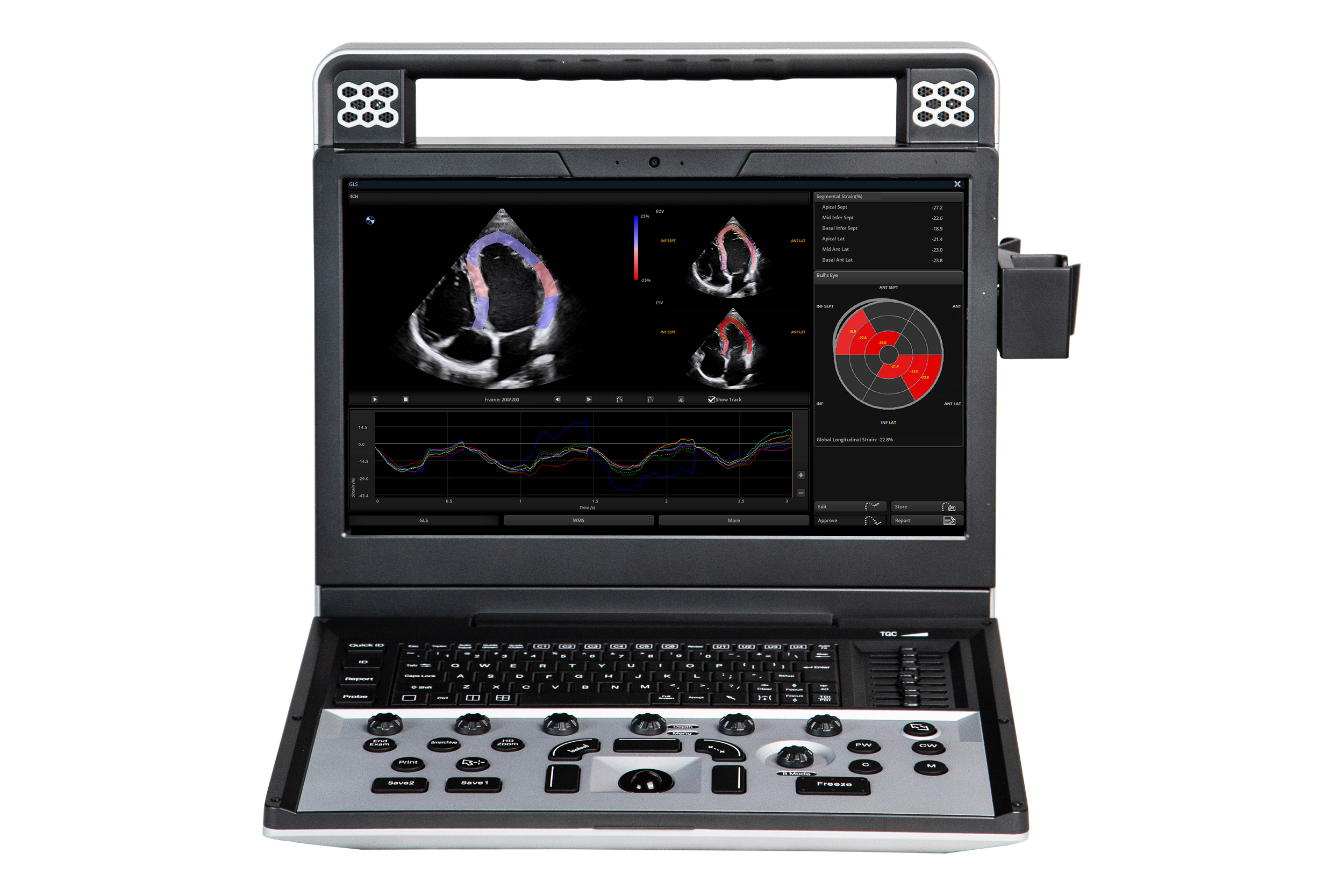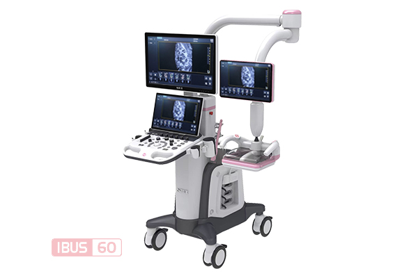
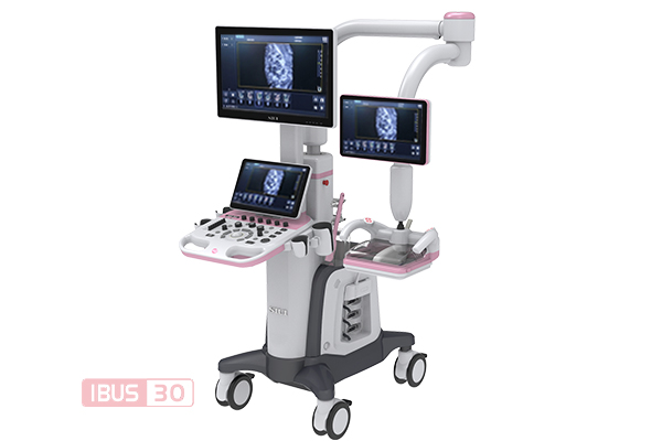
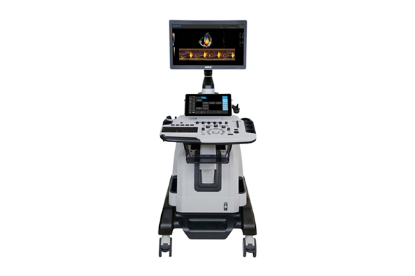
IBUS 60 breast ultrasound system
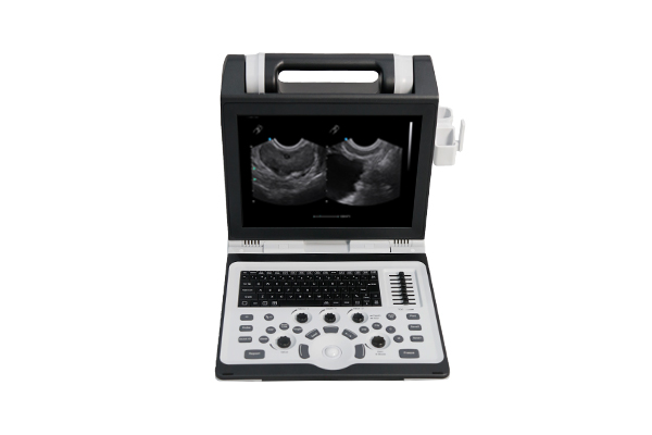
IBUS 30 breast ultrasound system
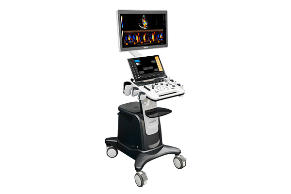
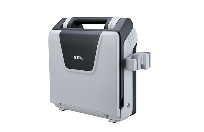
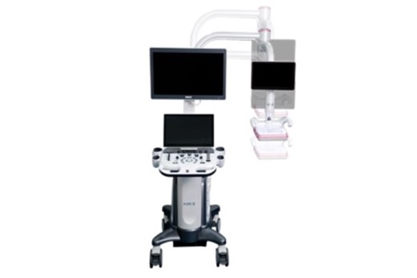
IBUS ultrasound system, rear view
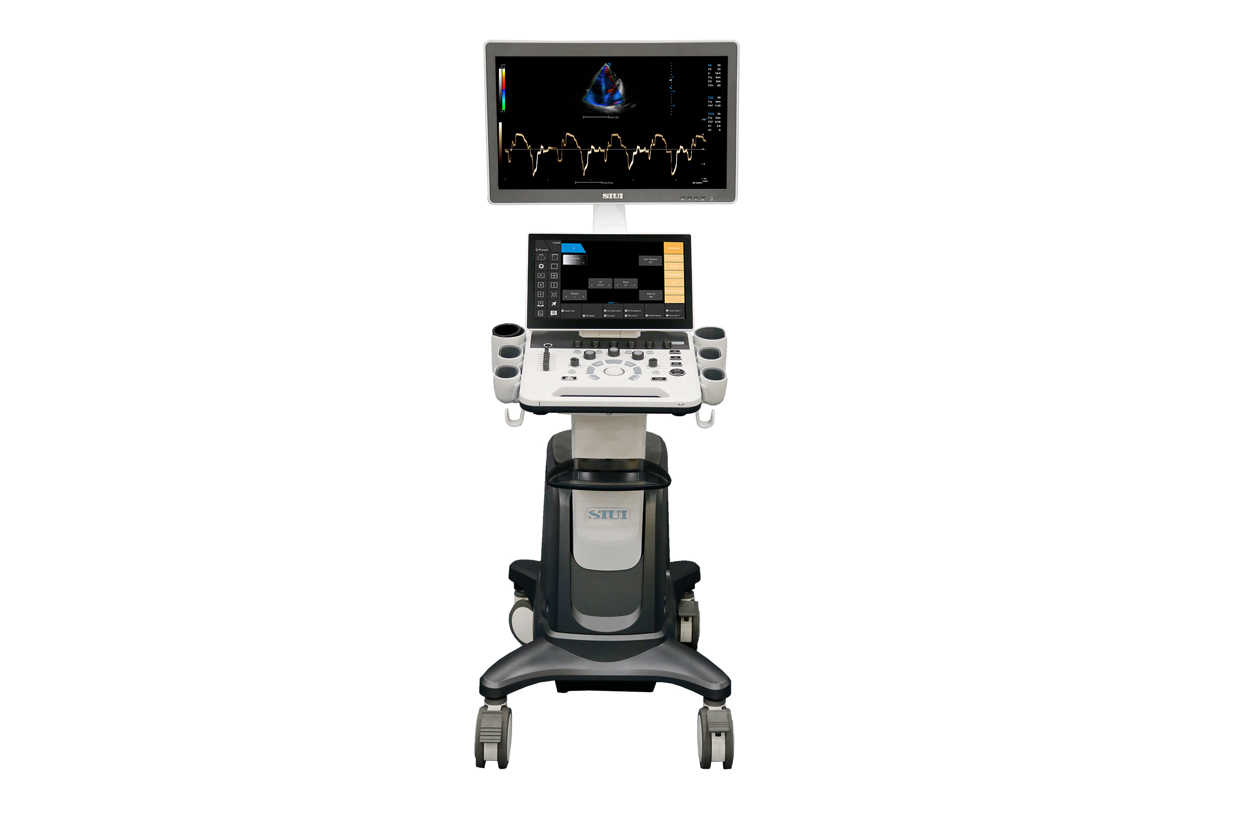
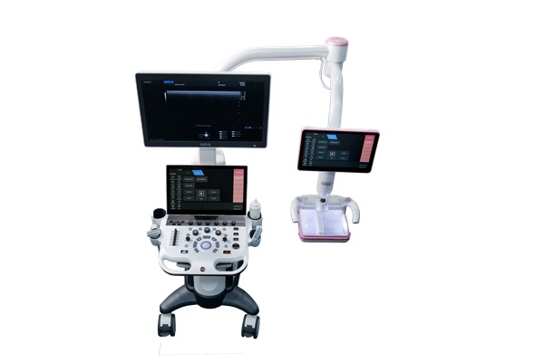
IBUS ultrasound system, front view
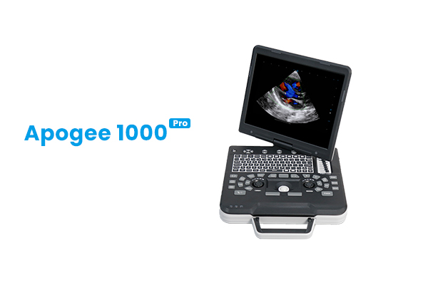
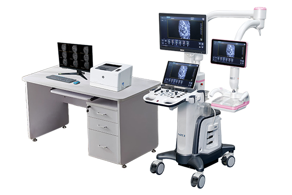
IBUS ultrasound system in a workstation setup
Human Ultrasound ยท Breast Volume Ultrasound

IBUS

Superb Versatility in Women's and Children's Healthcare

An intelligent breast volume diagnostic system with a specialised breast probe and standardised, BI-RADS-guided scanning, also covering OB/GYN and paediatric applications.

  • Specialised breast probe with full volume imaging
  • Standardised, BI-RADS-guided scanning workflow
  • OB/GYN and paediatric applications
Specifications
Overview

Superb Versatility in Women's and Children's Healthcare

IBUS is an intelligent breast volume diagnostic system for women’s and children’s healthcare. The series, IBUS 60 and IBUS 30, combines a specialised breast probe with a standardised scanning and diagnosis process guided by BI-RADS.

Beyond breast imaging, it offers OB/GYN and paediatric applications, including automated fetal measurements, 4D fetal imaging and infant craniocerebral and hip assessment.

Key Features

Designed around the clinician

The hardware and workflow features of IBUS, as published by the manufacturer.

Outlook Design on the IBUS

Outlook Design

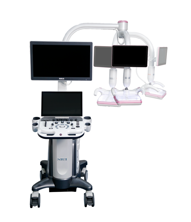
  • High-resolution monitor and dual touch screens.
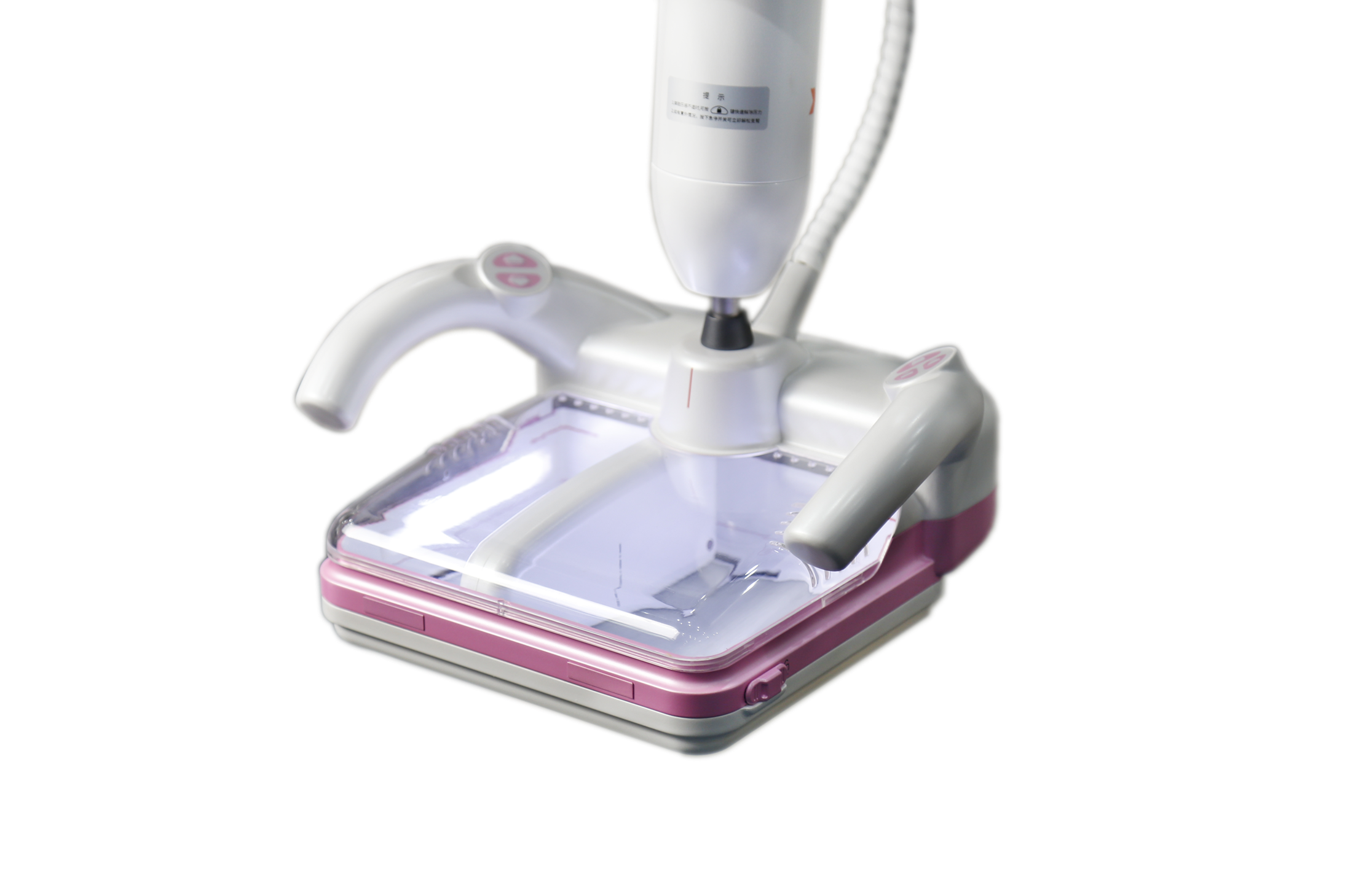
  • Specialised breast probe and 3 common probe connectors.
  • Large-sized breast probe for efficient scanning.
Low damping articulated arm on the IBUS

Low damping articulated arm

  • Low damping articulated arm with a wide range of adjustment.

Patented coupling film

  • Patented coupling film for enhanced patient comfort.

Standardized & Customizable Workflow

  • A standardised process helps avoid missed diagnosis.
  • Customisable scanning sections adapt to specific cases.

IBUS vs Traditional Ultrasound

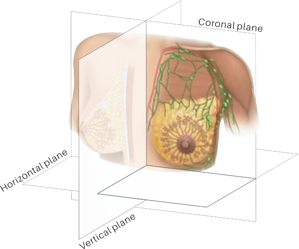
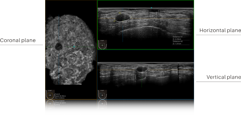
  • 3D imaging in coronal, transverse and sagittal planes.
  • Field of view more than 3 times larger than a traditional 2D probe.

IBUS vs Mammography

  • No radiation dose.
  • Better detection in dense breasts.

IBUS vs MRI

  • Shorter exam time.
  • Harmless for all people.
  • Cost-effective.
Clinical Functions

Clinical tools and applications

Automated measurement and imaging functions available on IBUS.

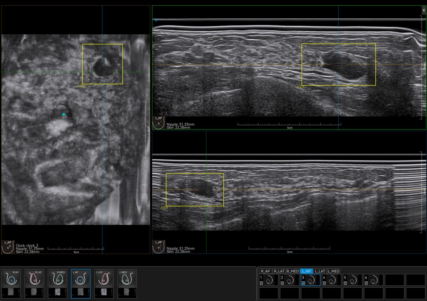
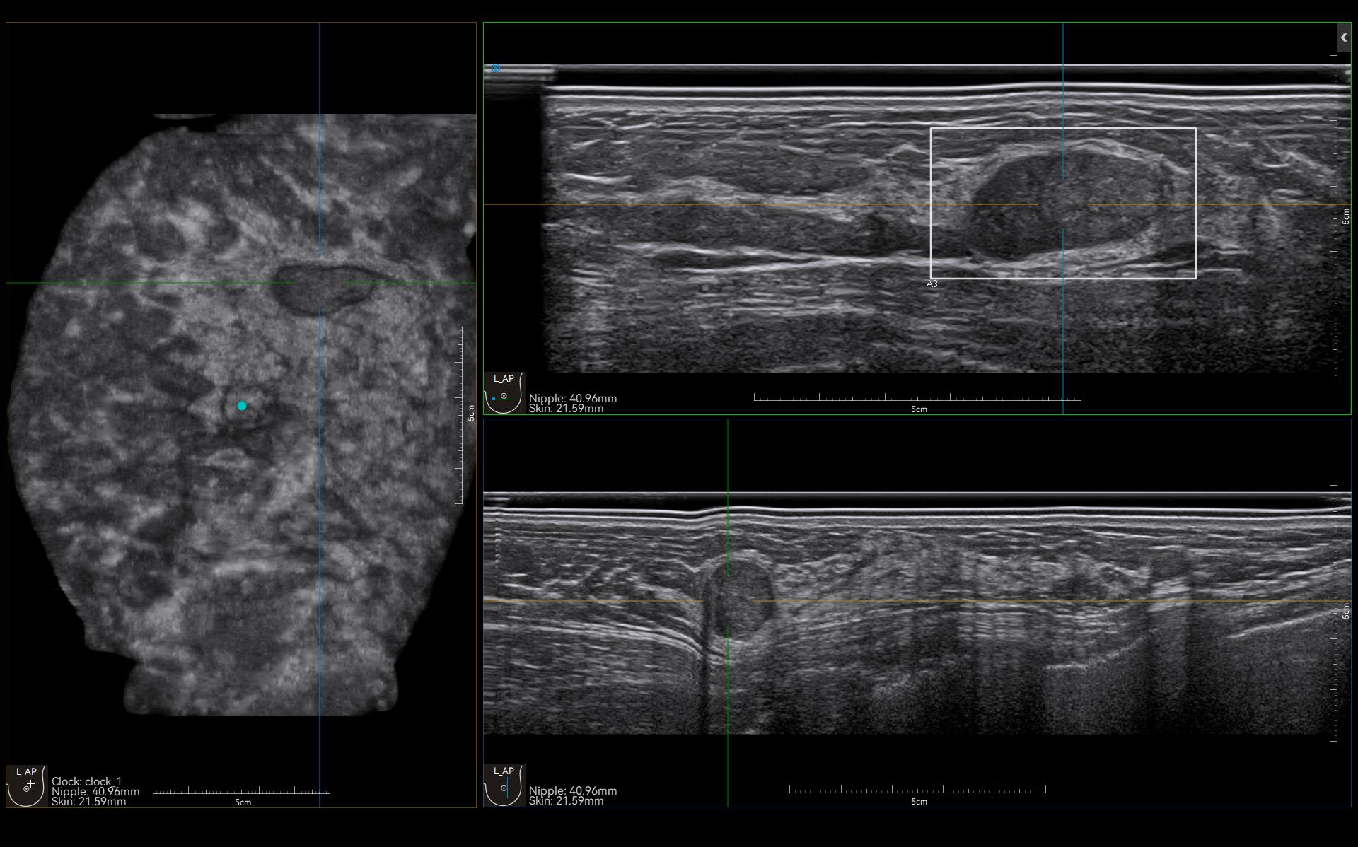
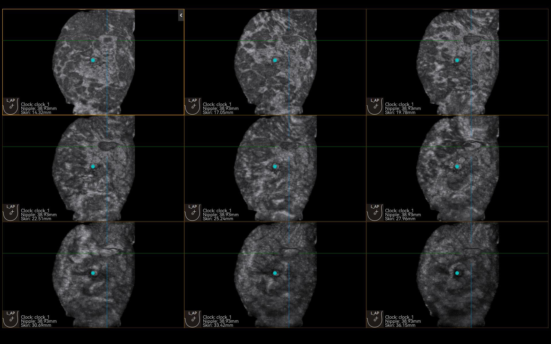
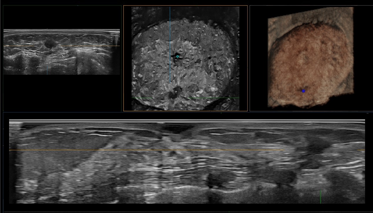
Full Volume Imaging and Coronal Section with High Clinical Value on the IBUS (image 1 of 2)Full Volume Imaging and Coronal Section with High Clinical Value on the IBUS (image 2 of 2)
01 ยท Breast

Full Volume Imaging and Coronal Section with High Clinical Value

Acquires volume data from multiple sections. The coronal view shows breast anatomy in the supine position to support surgical planning.

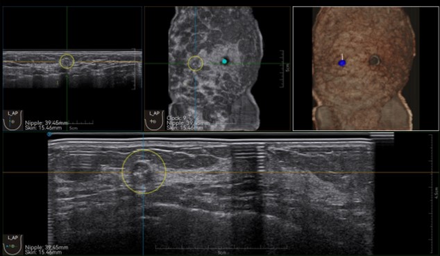
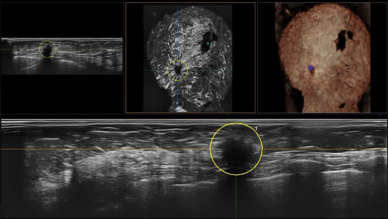
Automatic lesion detection on the IBUS
02 ยท Breast

Automatic lesion detection

AI-based detection flags the suspected area and traces the lesion across sections, with coordinate and graphic indication.

Lesion stereo images on the IBUS
03 ยท Breast

Lesion stereo images

Stereo images rendered from the volume data for lesion observation and localisation.

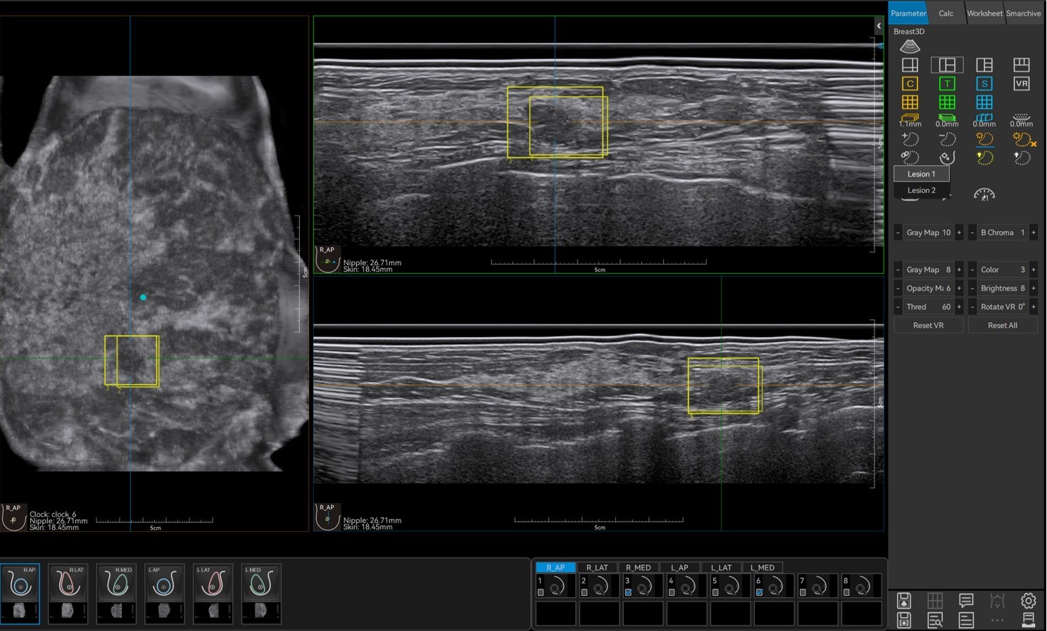
Lesion correlation on the IBUS
04 ยท Breast

Lesion correlation

Correlates information from different scan positions and sections to help avoid misdiagnosis.

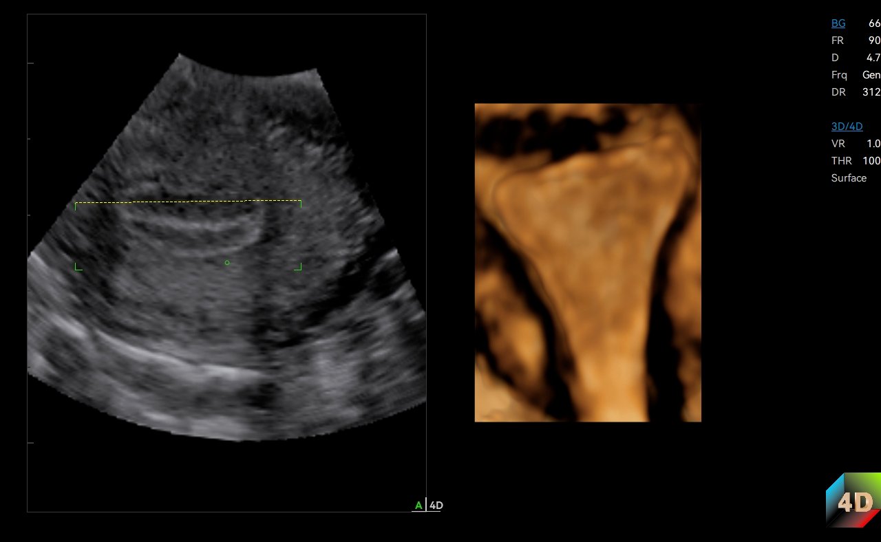
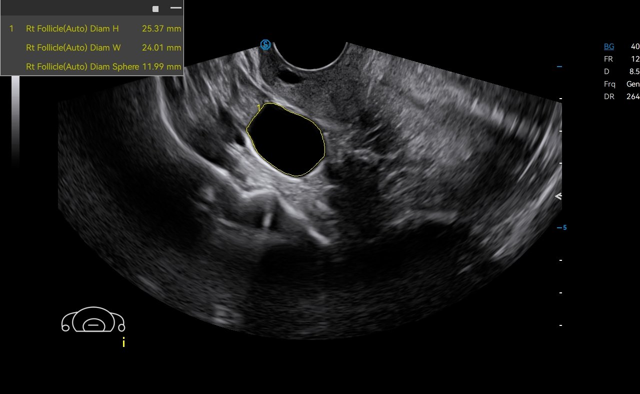
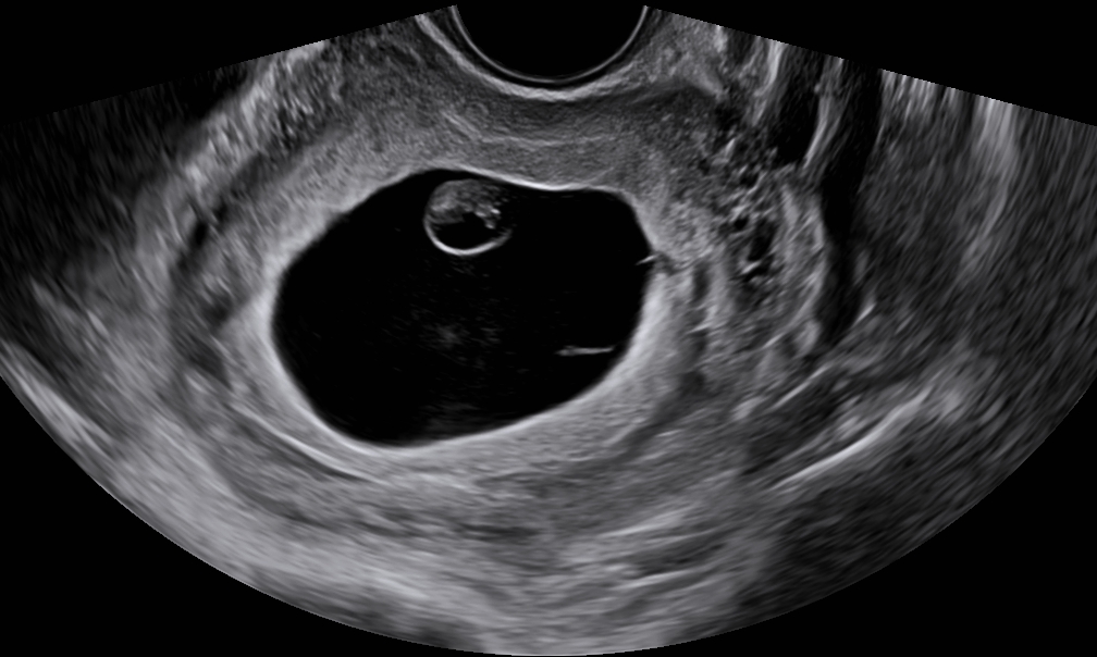
4D imaging on the IBUS
05 ยท OB/GYN

4D imaging

Uses a 4D vaginal probe to observe organs such as the uterus more closely.

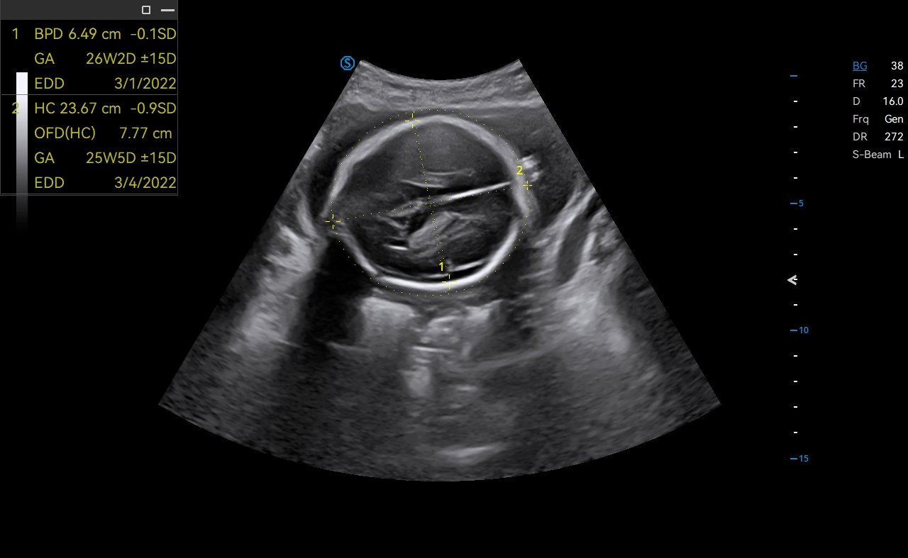
Auto OB on the IBUS
06 ยท OB/GYN

Auto OB

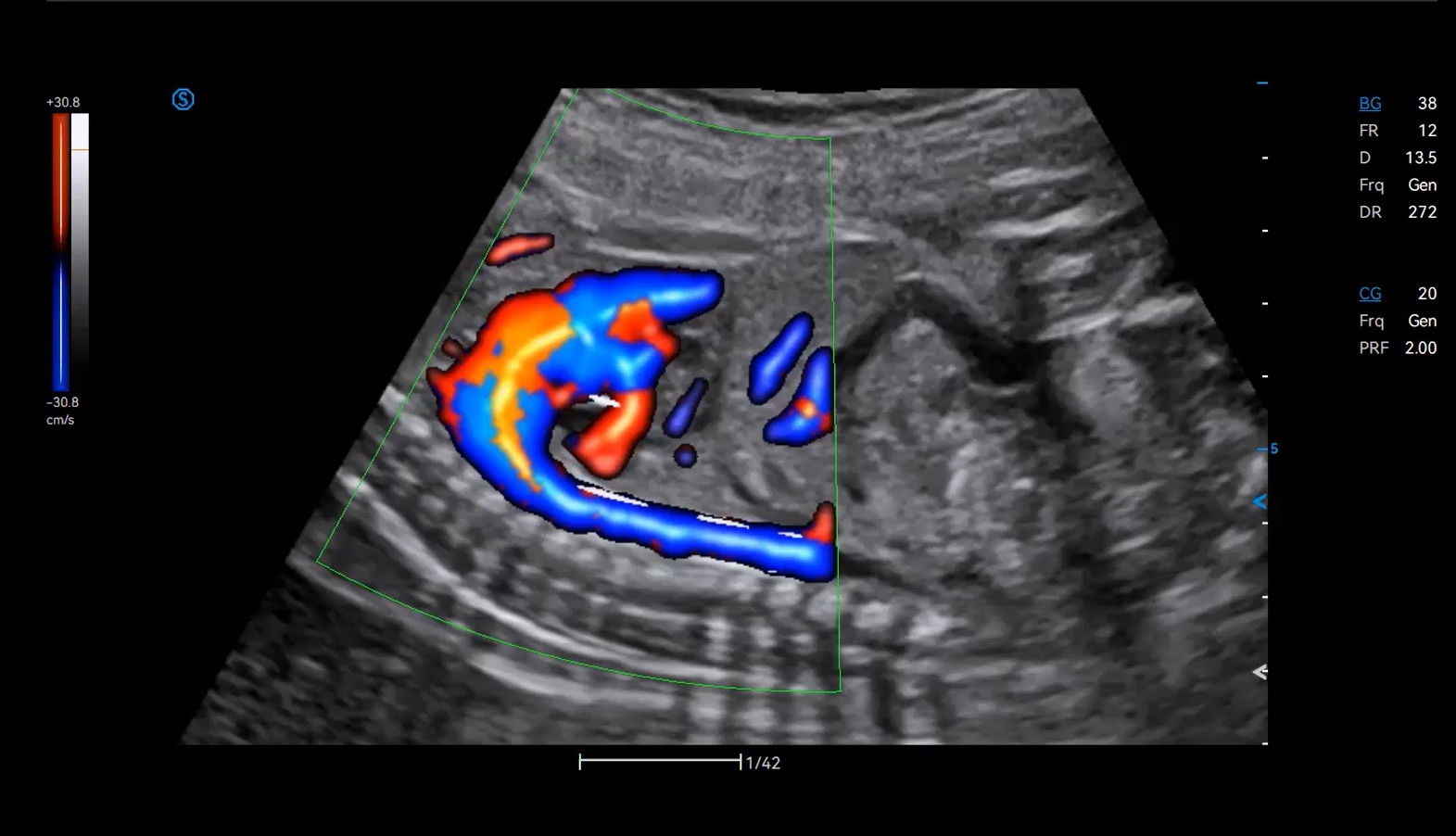
Automatically measures common fetal biometry (HC, BPD, FL and more) in standard sections.

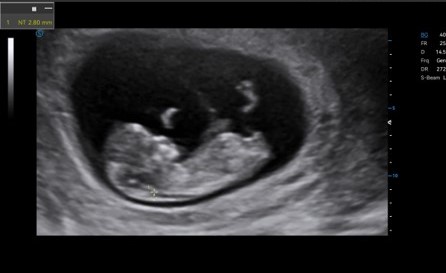
Auto NT on the IBUS
07 ยท OB/GYN

Auto NT

Recognises and measures fetal nuchal translucency thickness.

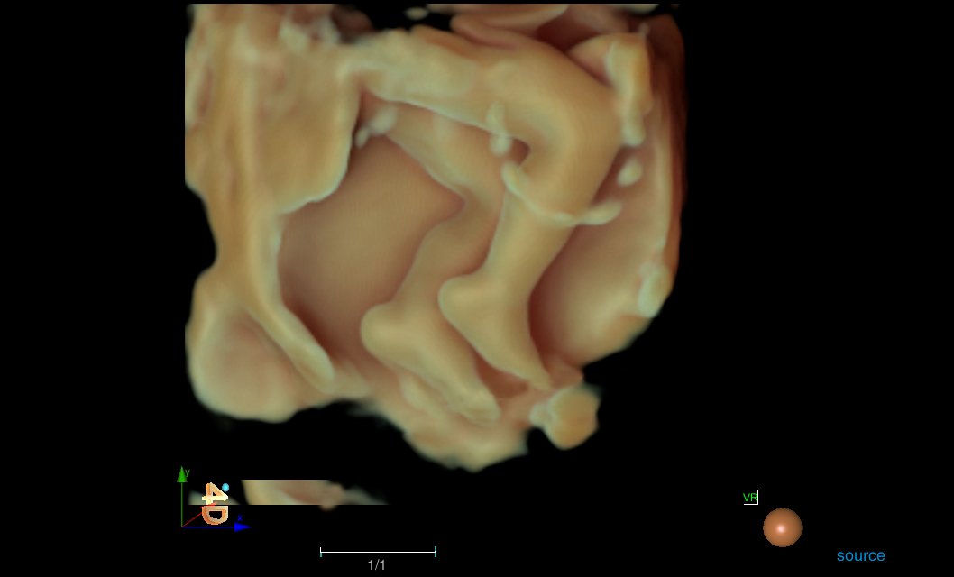
Lumi 4D on the IBUS
08 ยท OB/GYN

Lumi 4D

Adjustable light source angle for real-time static stereo imaging of the fetus.

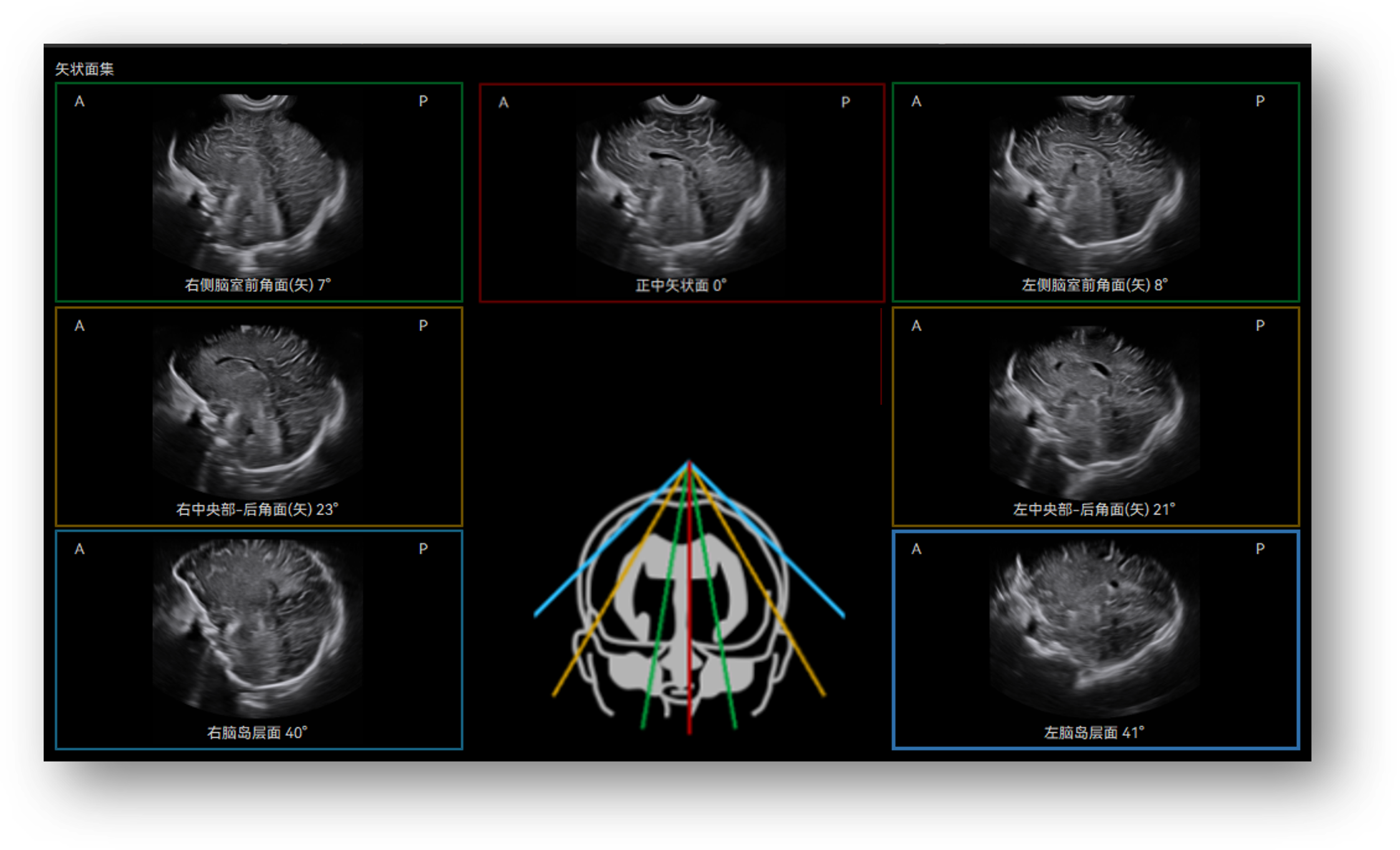
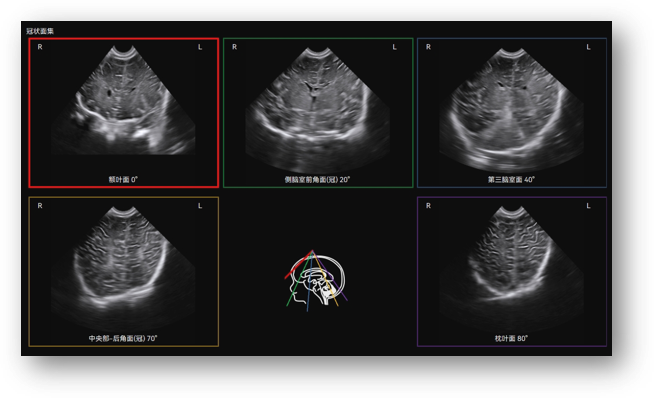
Craniocerebral Ultrasound Tomography (Infant) on the IBUS (image 1 of 2)Craniocerebral Ultrasound Tomography (Infant) on the IBUS (image 2 of 2)
09 ยท Paediatrics

Craniocerebral Ultrasound Tomography (Infant)

Identifies the 12 standard craniocerebral sections from volume data.

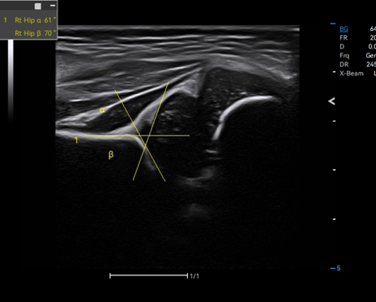
Auto Hip on the IBUS
10 ยท Paediatrics

Auto Hip

Identifies the baseline, tracks the bone and cartilage top line and measures for hip dysplasia evaluation.

Technical Specifications

IBUS at a glance

Specifications listed are those published by the manufacturer. For full technical documentation or a specific configuration, please contact our team.

Request full specifications
System Class
Specialised cart-based Breast Screening + OB/GYN + Pediatric solutions
Monitor / Display
23-inch high-resolution monitor + 15.6 inch touch touchscreen
Probe Connectors / Types
3 probe connectors
Imaging Modes
Breast Screening, OB/GYN and Pediatric solutions
Imaging Technologies
3D/4D imaging, auto lesion detection, strain elastography; 4D vaginal imaging, auto fetal measurements, Auto NT, Auto OB, Cerebral Tomograpy (infant), Auto-Hip
Connectivity / Other
Integrated gel warmer, offline workstation
Clinical Applications

Where IBUS is used

Breast clinics
Women's health centres
Pregnancy scans
Child health checks
Fertility clinics
Breast cancer screening
Product Enquiry

Enquire about IBUS

Ask about configurations, availability, demonstrations or pricing. Your enquiry is pre-filled for IBUS.

    IBUS enquiry

    Tell us about your practice and the equipment you are considering. Fields marked * are required.