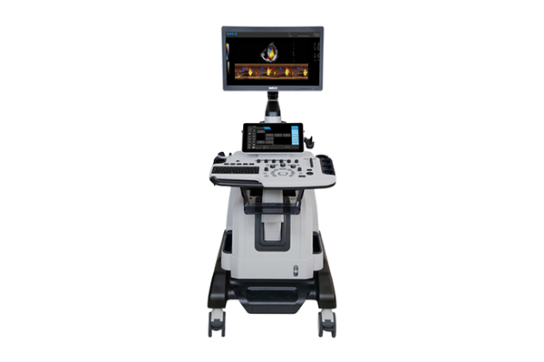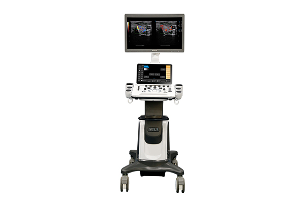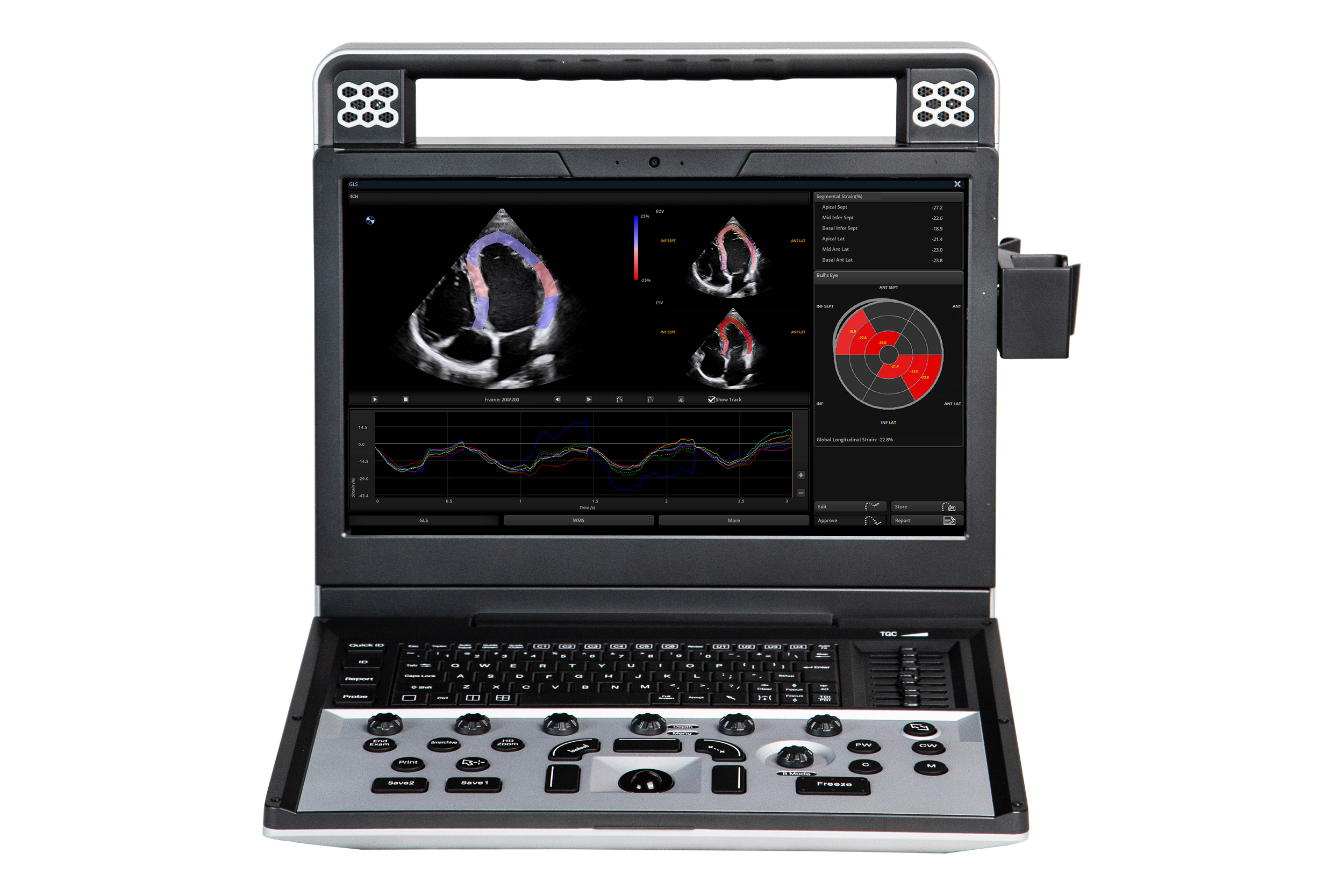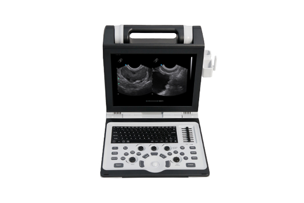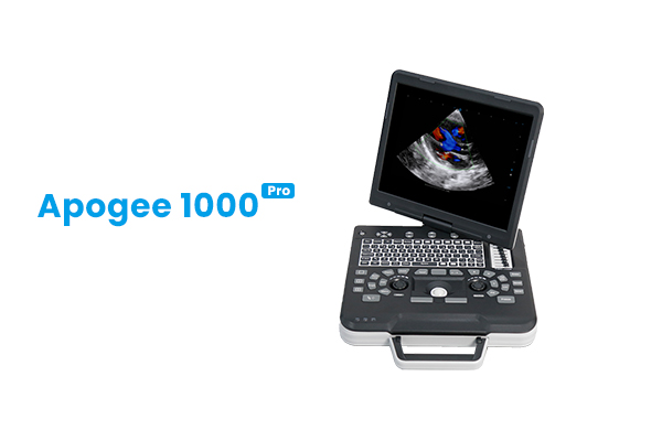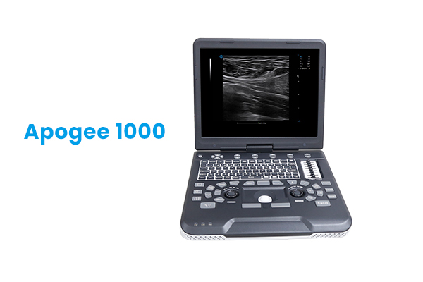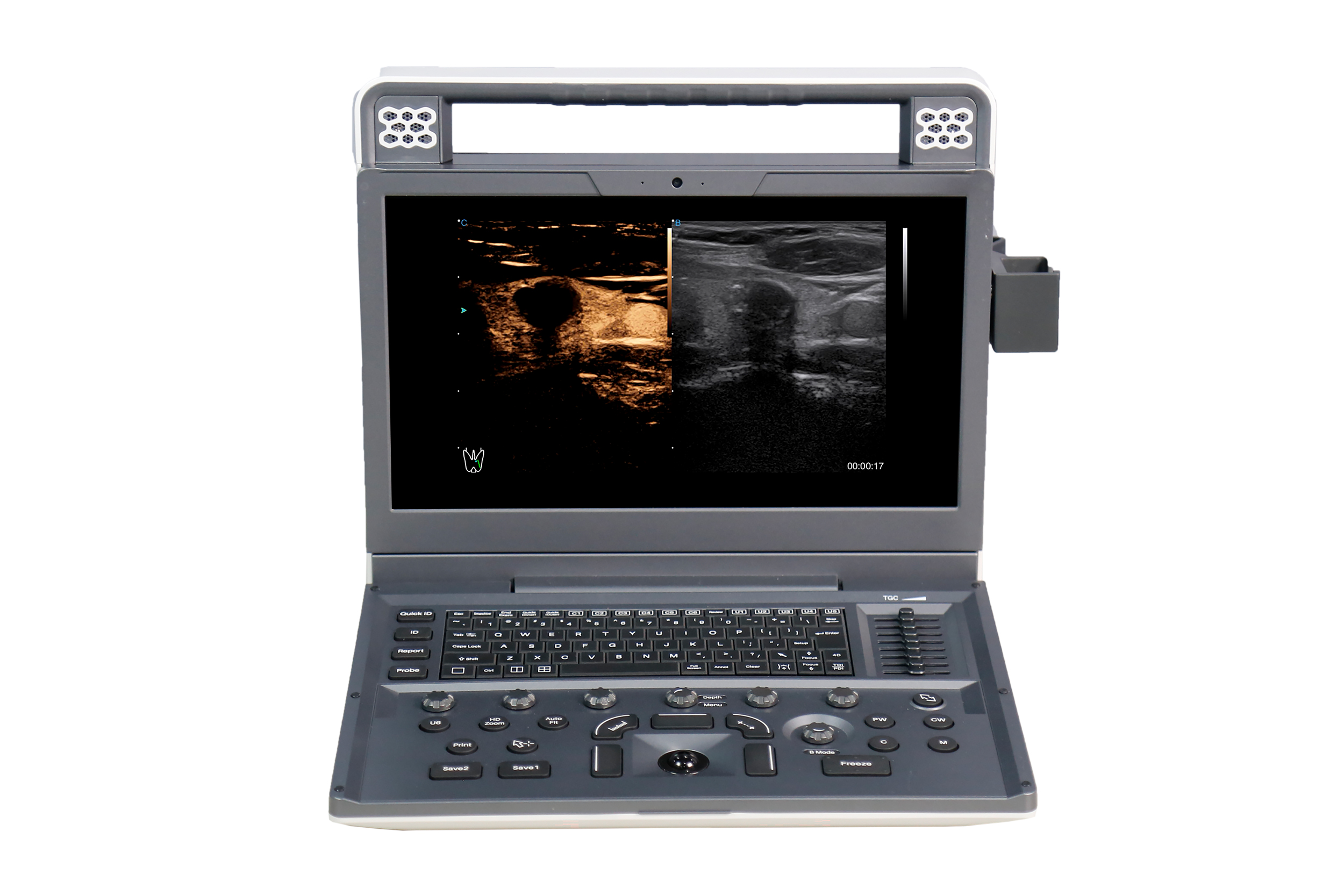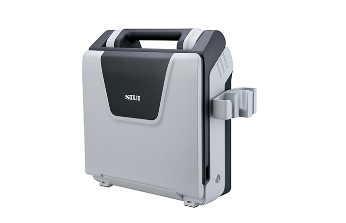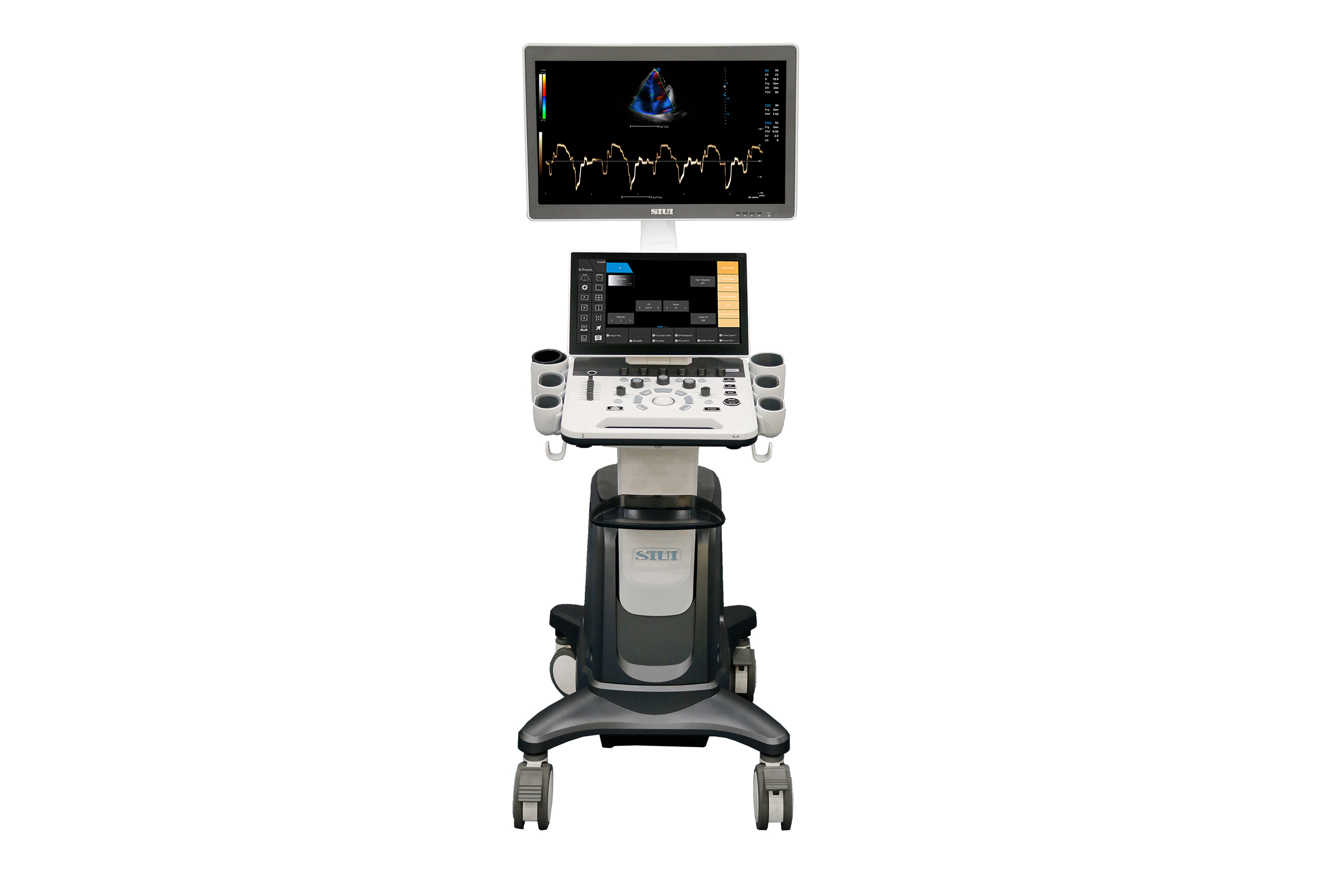
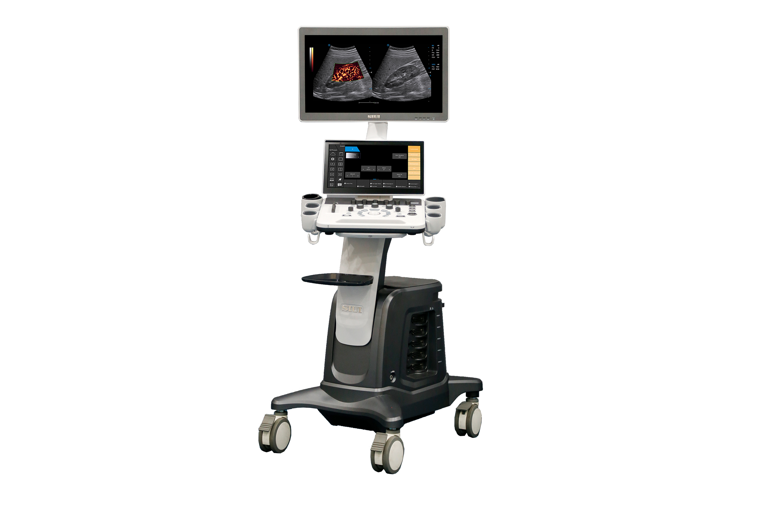
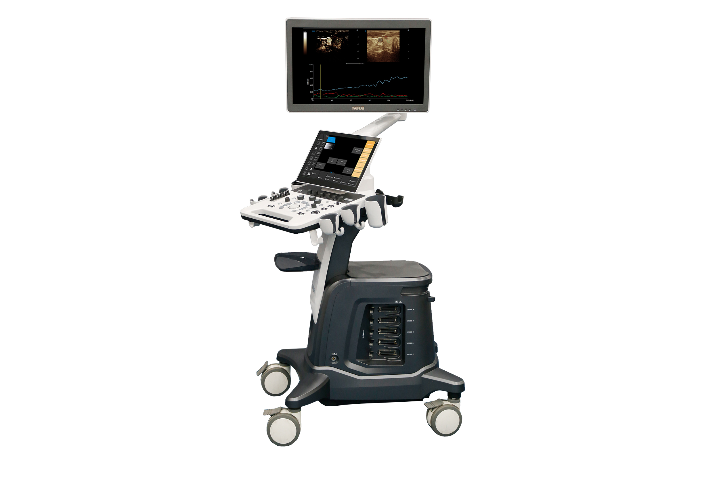
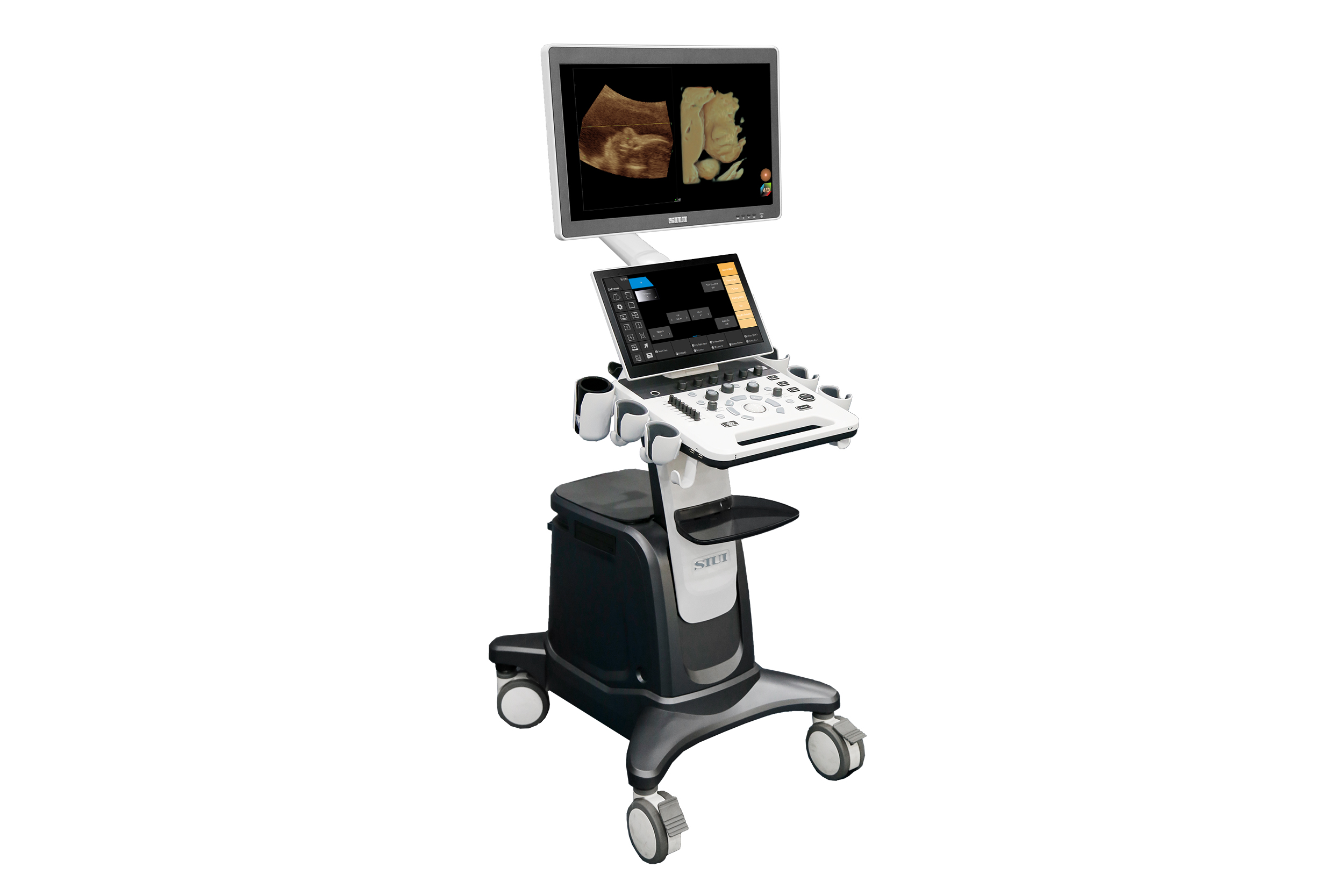
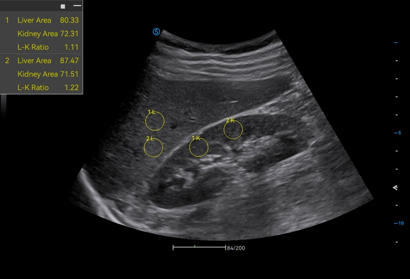
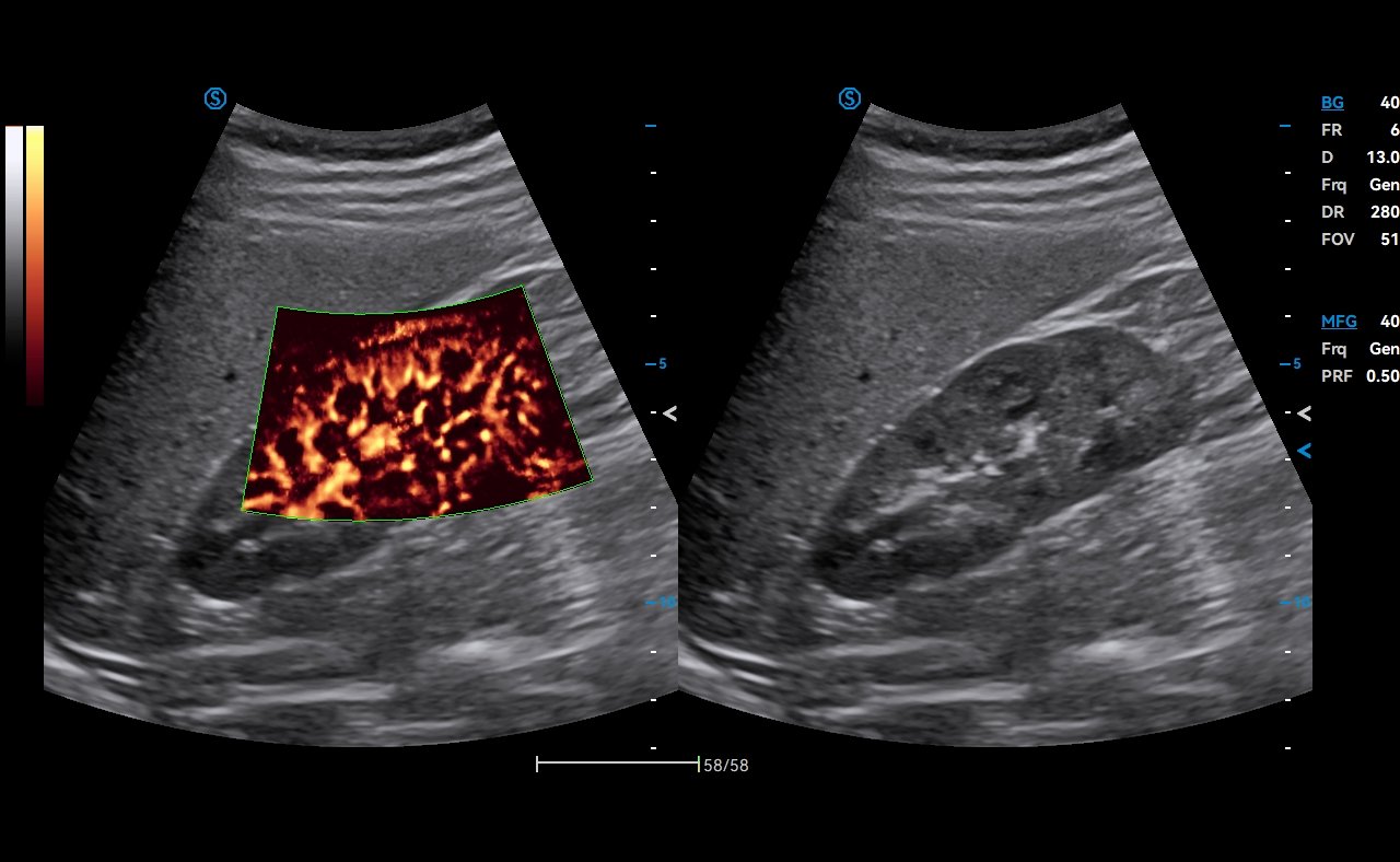
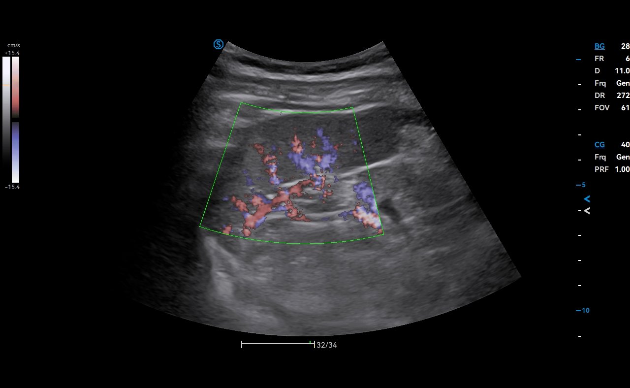
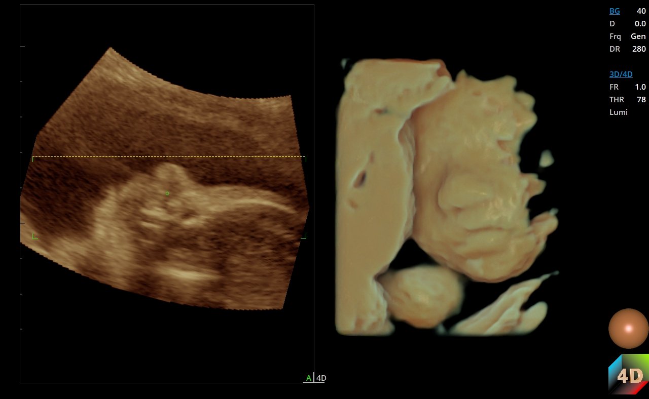
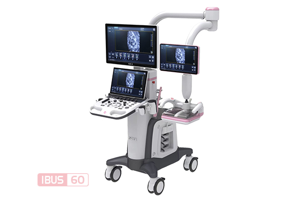
A50 ultrasound system
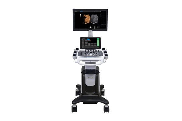
A50 ultrasound system with OB image on screen
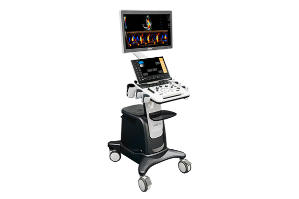
A50 ultrasound system with cardiac spectral Doppler on screen
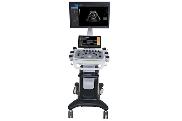
A50 ultrasound system with 4D image on screen
Human Ultrasound · Color Doppler

A50

Comprehensive imaging, intelligent cardiology

A versatile colour Doppler system covering a comprehensive range of applications from primary healthcare to advanced cardiology.

  • 21.5-inch adjustable monitor (upgradable to 23-inch)
  • 13.3-inch touch screen (upgradable to 15.6-inch)
  • 5 activated probe connectors
Specifications
Overview

Comprehensive imaging, intelligent cardiology

A50 is an outstanding solution covering a comprehensive range of applications from basic to advanced levels, addressing diverse needs of patients across all ages and genders in primary healthcare.

It also delivers intelligent solutions for cardiology to enhance diagnostic confidence for clinicians.

Key Features

Designed around the clinician

The hardware and workflow features of A50, as published by the manufacturer.

Elegant Design

  • 21.5-inch adjustable monitor (upgradable to 23-inch)
  • 13.3-inch touch screen (upgradable to 15.6-inch)
  • 5 activated probe connectors
  • 6 stable probe holders
  • Dedicated holder for intracavity probe
  • Built-in batteries for extended use (optional)

Compatible with Diverse Probes

  • Linear probe
  • Phased array probe
  • Convex probe
  • Micro-convex probe
  • Micro-convex volumetric probe
  • 4D convex probe
  • Transvaginal probe
  • TEE probe
Clinical Functions

Clinical tools and applications

Automated measurement and imaging functions available on A50.

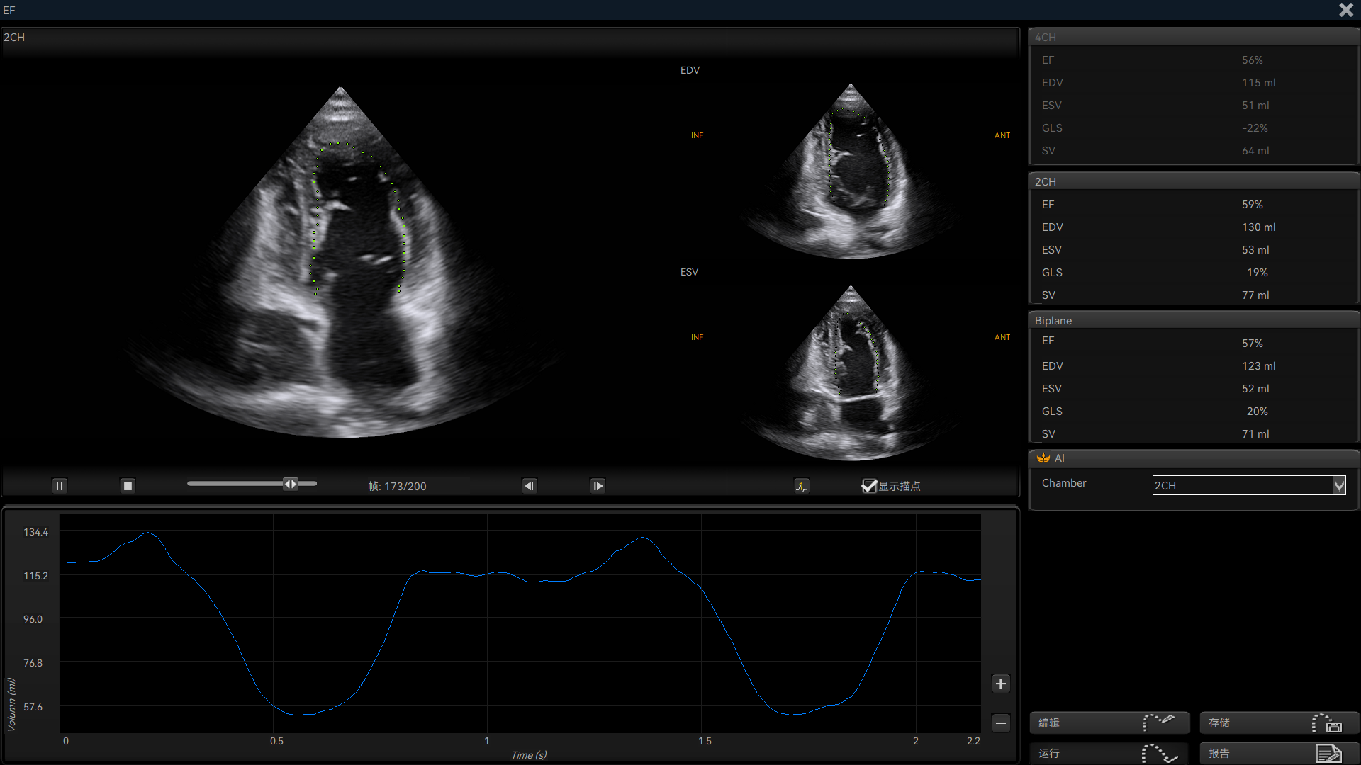
Auto EF Plus on the A50
01 · Cardiology

Auto EF Plus

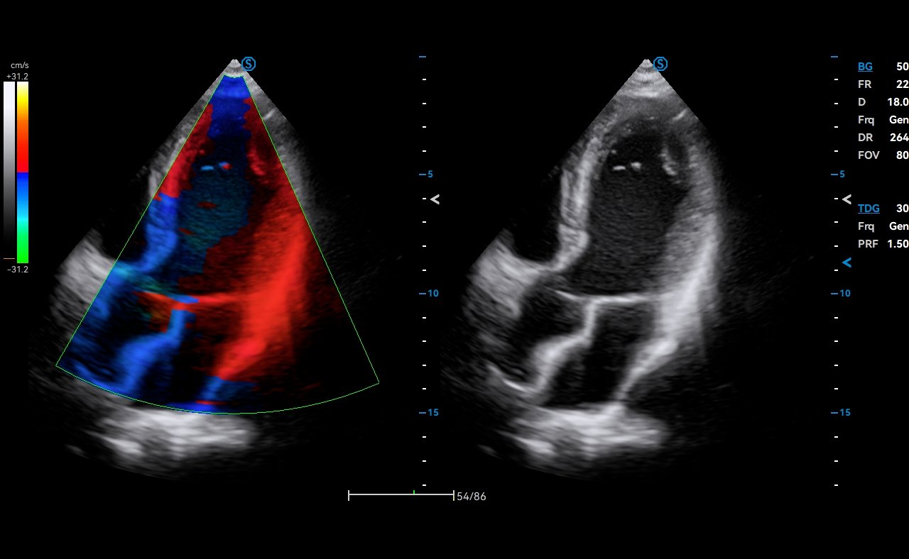
Automatically tracks the endocardium and calculates LV EF within seconds for systolic function assessment. AI-powered cardiac section identification simplifies the operation.

Auto SG Plus on the A50
02 · Cardiology

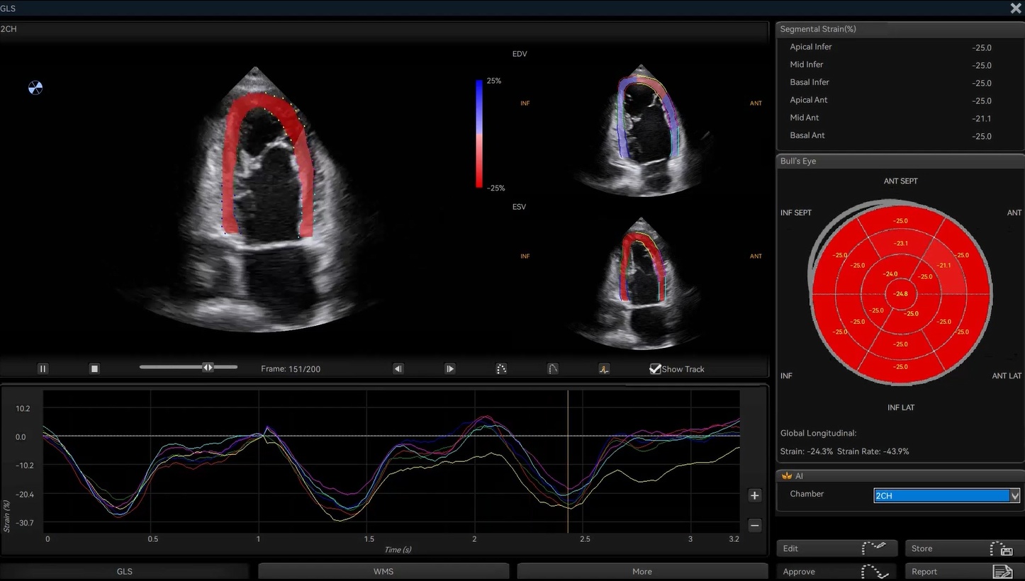
Auto SG Plus

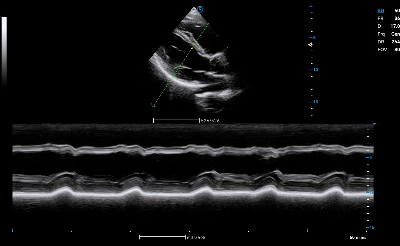
Quantifies global and segmental LV function in a Bull's-eye diagram, while AI-powered cardiac plane identification simplifies operation.

Craniocerebral Ultrasound Tomography (Infant) on the A50 (image 1 of 2)Craniocerebral Ultrasound Tomography (Infant) on the A50 (image 2 of 2)
03 · Abdomen

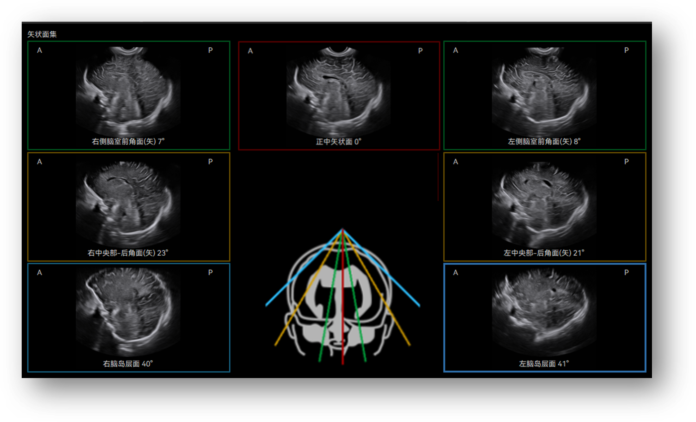
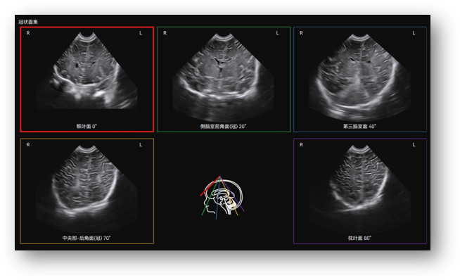
Craniocerebral Ultrasound Tomography (Infant)

Automatically identifies and displays the 12 standard craniocerebral sections using a micro-convex volumetric probe, enabling efficient evaluation of infant craniocerebral development.

CHI on the A50 (image 1 of 2)CHI on the A50 (image 2 of 2)
04 · Abdomen

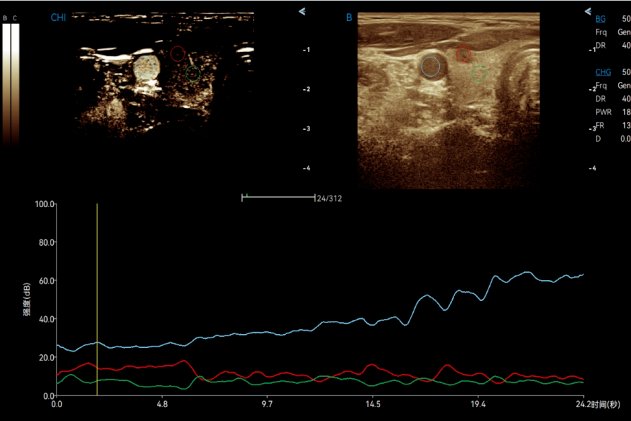
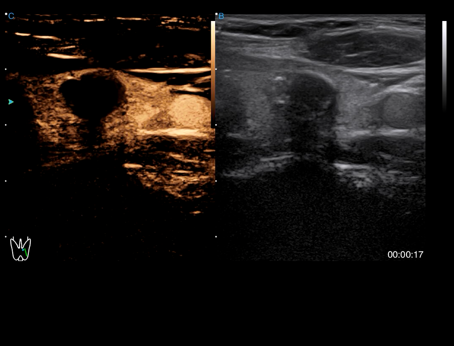
CHI

Enables real-time visualization of blood perfusion status of the organs (liver, thyroid, breast) with enhanced contrast, while CHI QA offers quantitative results for improved precision.

Strain Elastography on the A50
05 · Elastography

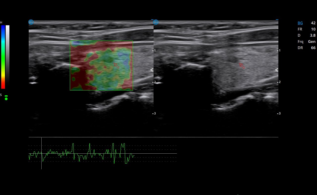
Strain Elastography

Visualizes the stiffness of tissues in real time to detect potential abnormalities.

Technical Specifications

A50 at a glance

Specifications listed are those published by the manufacturer. For full technical documentation or a specific configuration, please contact our team.

Request full specifications
System Class
High-performance, cart-based
Monitor / Display
21.5-inch + 13.3-inch touchscreen
Probe Connectors / Types
5
Imaging Modes
B, M/AMM, CFM/PDI/DPDI/VS Flow, PW/HPRF/CW, TDI, 3D/4D
Imaging Technologies
THI, MFI, X-Beam, Nanoview/Nanopure, Strain Elastography, Needle Enhancement
Clinical Applications

Where A50 is used

Flagship hospital imaging
High-volume clinics
Biopsy procedures
Heart and vascular scans
Teaching hospitals
Multi-department imaging
Product Enquiry

Enquire about A50

Ask about configurations, availability, demonstrations or pricing. Your enquiry is pre-filled for A50.

    A50 enquiry

    Tell us about your practice and the equipment you are considering. Fields marked * are required.