Apogee C5
- Details
- Category: Color Doppler

Features
Apogee C5
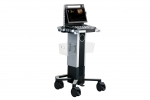
Apogee C5 is an exceptional integration of charming outlook, considerate workflow, clever imaging processing platform, and outstanding-performance images. In addition to general examinations, this portable ultrasound system is capable of offering cutting-edge application solutions in cardiology and pediatrics to enhance physicians’ diagnostic confidence.
Charming Design with Exquisite Aesthetic
● 15.6-inch adjustable monitor
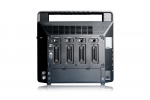
● 4 probe connectors
● Digital power indicator
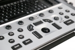
● User-centered keyboard layout
Compatible with trolley CR-50
● Adjustable height
● 6 stable probe holders
● Front and rear cart handles for easy movement
Clever Realview+ Platform
● Pixel Echo Zone present images at high frame rate through wide-band information processing while maintaining consistent focus.
● Tailored Filter extracts effective information and filters noise to achieves high image contrast.
● S-Beam avoids image distortion to improve overall fidelity by compounded compensation.
Considerate user-oriented workflow
● Quick ID: one-touch quick ID creation
● S-view: simultaneously compare multiple images and cines
● Historical file query: automatically retrieves relevant data from previous exams when entering the existing patient ID
● Customized report template and exam type: adapt to diverse usage habits
● Q-Preset: self-defined parameters optimize application efficiency
Click on images to enlarge
Functions
Cutting-Edge Application Solutions Enhance Diagnostic Confidence
Auto EF facilitates cardiac systolic function assessment by automatically identifying and tracking the endocardium, and accurately calculating the LV EF within seconds.

Auto SG empowers objective evaluation of LV systolic function and myocardial deformation through quantitative results in a bull’s-eye plot.

TDI QA quantitatively analyzes the myocardial motion with multiple sampling points for convenient comparison and evaluation.

Stress Echo analyzes myocardial motion at rest and under stress to help evaluate how coronary arteries respond to the stress.

CHI QA visualizes the organ blood perfusion status with enhanced contrast. Dual mode comparison and time-intensity curves provide objective results for tumor diagnosis.

Strain Elastography distinguishes the stiffness levels of tissues in real time to detect potential abnormalities.

Micro Flow sensitively detects micro blood flow signals to precisely reflect the blood flow perfusion status in organs.

Competitive Pediatrics Application Solutions
Craniocerebral Ultrasound Tomography (Infant) significantly simplifies the examination by automatically identifying and displaying the 12 standard craniocerebral sections.

Professional Probes for Paediatric Cardiac Examinations
Micro-Convex Probe - R11 (C6)
Phased Array Probe - P8
Phased Array Probe - P5
Pediatric TEE Probe
 |
 |
● Dedicated for premature infant, newborn and child
● Small footprint design
● High frequency and high resolution


